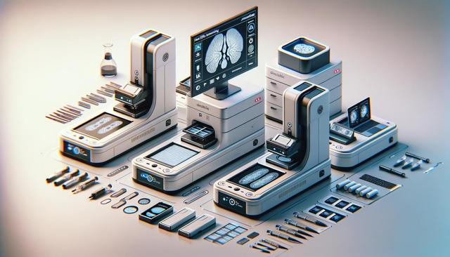
Advancing Histopathology: Digital Slide Scanners with AI and Secure Data Storage
Digital Histology and the Role of Whole Slide Imaging
Whole Slide Imaging (WSI) technology is reshaping how histological samples are analyzed, shared, and archived. Traditionally, pathologists relied on optical microscopes to examine glass slides, which posed challenges in terms of accessibility, reproducibility, and data sharing. With the rise of digital histology, pathology slide scanners now convert entire glass slides into high-resolution digital images. These digital scans provide a comprehensive view of tissue samples, enabling clinicians and researchers to evaluate them on-screen with exceptional clarity.
WSI devices are equipped with advanced optics and motorized scanning stages that ensure consistent, high-quality image capture. These scanners can handle different magnifications and staining techniques, accommodating a range of diagnostic needs. By digitizing slides, institutions benefit from easier storage, faster retrieval, and remote access, all of which support a more agile and collaborative workflow.
AI Annotation: Enhancing Diagnostic Accuracy and Efficiency
One of the most impactful developments in digital pathology is the integration of artificial intelligence (AI) into slide analysis. Modern pathology slide scanners now come with software capabilities that leverage AI for automated annotation, pattern recognition, and quantification. These tools assist pathologists by identifying regions of interest, highlighting abnormal structures, and suggesting potential diagnoses based on trained algorithms.
AI-powered annotation offers several advantages:
- Reduces human error through consistent interpretation of complex patterns
- Speeds up the diagnostic process by pre-screening slides
- Supports educational and training efforts with standardized references
While AI does not replace the role of expert pathologists, it serves as a valuable tool that enhances their decision-making, particularly in high-volume or complex cases. As machine learning models improve, their reliability and utility in clinical settings will likely expand.
Secure Data Storage Solutions for Digital Pathology
With the transition to digital formats, data security and storage have become critical components of pathology workflows. Whole slide images are large files, often several gigabytes in size, necessitating robust storage infrastructure. Moreover, these images are part of patient medical records and must meet stringent privacy and compliance standards.
Modern pathology slide scanners are integrated with secure data storage solutions that offer:
- Encryption for data in transit and at rest
- Redundant backups to prevent data loss
- Compliance with health data regulations such as HIPAA or GDPR
- User authentication and role-based access control
Cloud-based storage platforms are increasingly used, offering scalability and remote access capabilities. Hybrid models, combining on-premise and cloud storage, are also common, providing flexibility and control over sensitive data.
Applications Across Research, Education, and Clinical Practice
Digital histology with AI annotation and secure storage is not limited to clinical diagnostics. Its applications extend into academic research, pharmaceutical development, and medical education. For researchers, digital slides enable precise, reproducible analysis and easier collaboration across institutions. They can annotate, share, and compare slides without physical limitations, accelerating the pace of discovery.
In education, digital slides are replacing traditional teaching sets. Students can access a vast library of annotated images, practice diagnostic skills, and receive feedback through AI-supported learning platforms. This approach caters to diverse learning styles and supports remote or hybrid education models.
Clinically, digital slide scanners are especially valuable in remote or underserved areas where access to specialized pathologists is limited. Telepathology enables real-time consultation and second opinions, improving patient outcomes by connecting local facilities with global expertise.
Considerations for Implementing a Digital Pathology Workflow
Adopting pathology slide scanners and transitioning to digital histology requires thoughtful planning and investment. Institutions must evaluate factors such as scanner throughput, compatibility with existing lab systems, and integration with electronic medical records. Additionally, training staff to use new software tools, including AI-assisted annotation platforms, is essential to maximize their benefits.
Key considerations include:
- Scanner resolution, speed, and slide capacity
- AI software accuracy, usability, and customization options
- Data storage architecture and compliance readiness
- IT infrastructure for network performance and digital security
By addressing these areas, healthcare and research institutions can establish a reliable and scalable digital pathology environment that supports both current needs and future growth.
Conclusion: Empowering the Future of Pathology
Pathology slide scanners with whole slide imaging, AI annotation, and secure data storage are driving a significant shift in histological analysis. These technologies enhance diagnostic accuracy, streamline workflows, and broaden access to expert pathology services. Whether in clinical practice, research, or education, digital histology offers a powerful platform for innovation and collaboration. As adoption grows, institutions that invest in robust digital pathology solutions will be well-positioned to advance patient care and scientific discovery.


