Advancing Precision: Digital Microscopy Systems for High-Resolution Imaging
Understanding Digital Microscopy Systems
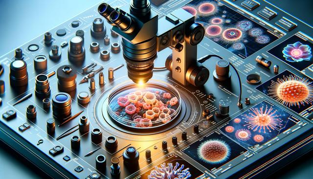
Digital microscopy systems represent a significant evolution from traditional optical microscopes by integrating high-performance cameras with computing power and image processing capabilities. These systems capture detailed digital images of microscopic samples, enabling enhanced analysis, storage, and sharing. Unlike conventional microscopy, digital systems offer features such as automated focus, image stitching, and high dynamic range imaging, which contribute to greater clarity and reproducibility in research settings. This technological shift is particularly beneficial in fields like biomedical research, materials science, and industrial inspections, where precision and documentation are critical.
At the core of these systems are lab-grade cameras that deliver high-resolution imaging with impressive clarity. These cameras often come equipped with CMOS or CCD sensors capable of capturing fine details even in low-light conditions. Paired with powerful software, they allow users to adjust parameters such as contrast, brightness, and magnification digitally, which facilitates deeper insights into sample morphology and structure. The result is not only improved image quality but also the ability to perform detailed quantitative analysis that would be difficult with analog methods.
Lab-Grade Cameras: Enhancing Image Clarity and Detail
The camera component in a digital microscopy system is crucial for achieving high-resolution imaging. Lab-grade cameras are engineered for scientific and industrial applications, ensuring consistent performance and accuracy. These cameras offer a range of features tailored to demanding environments, including:
- High megapixel counts for capturing fine structural details
- Fast frame rates for dynamic processes or live samples
- Low noise and high sensitivity for clear imaging in varied lighting conditions
- Compatibility with various microscope models through standardized mounts
Such capabilities make lab-grade cameras a valuable asset in pathology, semiconductor inspection, and forensic analysis. Their precision ensures that even minute features are visible, allowing researchers to identify patterns or abnormalities that might otherwise go unnoticed. Furthermore, advancements in sensor technology continue to push the boundaries of what is possible in digital imaging, expanding the application potential of these systems.
Advanced Analysis Software: From Observation to Insight
Equally important to the camera is the analysis software that powers digital microscopy systems. This software acts as the interface between the user and the imaging hardware, offering a suite of tools for image capture, processing, and interpretation. Its features typically include:
- Automated measurement tools for distance, area, and angle calculations
- Image enhancement algorithms to improve clarity
- Annotation and metadata tagging for contextual understanding
- Time-lapse and multi-channel imaging support
By integrating these functions, analysis software transforms raw image data into actionable insights. Researchers can compare samples over time, quantify changes, and generate comprehensive reports. Additionally, many software platforms now include AI-assisted analysis, which can identify cellular features or defects automatically, reducing the manual workload and increasing consistency. As a result, digital microscopy becomes not just a tool for observation but a full-fledged analytical platform.
Real-Time Collaboration: Connecting Teams Across Distances
One of the standout features of modern digital microscopy systems is their capability for real-time collaboration. By leveraging cloud connectivity and remote access, teams can view, discuss, and analyze samples simultaneously, regardless of their physical location. This functionality is particularly valuable in global research projects, academic instruction, and cross-site industrial inspections. Real-time collaboration tools include:
- Live image streaming with interactive controls
- Shared workspaces for annotations and data exchange
- Integrated video conferencing for seamless communication
- Cloud storage options for secure data access and backup
This collaborative environment fosters faster decision-making and enhances knowledge sharing. In educational settings, instructors can guide students through live demonstrations, while in the clinical context, specialists can consult on diagnoses without the need for physical sample transfers. The result is a more agile and connected research ecosystem where expertise can be accessed instantly.
Applications Across Diverse Fields
Digital microscopy systems are increasingly being adopted in a variety of disciplines due to their adaptability and precision. In life sciences, they are used for studying tissue samples, cellular behavior, and microorganism activity with unparalleled detail. In materials science, these systems help analyze surface structures, detect microfractures, and assess material integrity. Other areas benefiting from digital microscopy include:
- Pharmaceutical research for drug development and quality control
- Electronics and semiconductor industries for inspecting microcircuits
- Environmental sciences for analyzing soil, water, and air particulates
- Forensic science for evidence examination
The versatility of digital microscopy systems, combined with their ability to deliver high-resolution imaging and facilitate real-time collaboration, makes them a valuable tool across these sectors. Their continued development is expected to unlock new possibilities in both research and applied sciences.
Conclusion: Empowering Precision and Collaboration
Digital microscopy systems are redefining how researchers and professionals capture, analyze, and share microscopic data. By integrating lab-grade cameras, sophisticated analysis software, and real-time collaboration tools, these systems provide a comprehensive solution for high-resolution imaging. Whether in a laboratory, industrial, or educational setting, they support greater accuracy, efficiency, and connectivity. As the technology continues to evolve, digital microscopy is becoming an indispensable asset for those seeking deeper insights and improved workflows in a wide range of scientific and technical fields.


