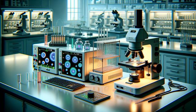
Advancing Research with Live Cell Imaging: Tools for Real-Time Insight
Understanding Live Cell Imaging and Its Importance
Live cell imaging is a vital technique in modern biological and biomedical research. It allows scientists to observe live cells in real time, providing insights into cellular behavior, interactions, and responses to various stimuli. This method is particularly useful for studying dynamic processes like cell division, migration, and apoptosis. Unlike fixed cell imaging, live cell analysis preserves the natural physiological state of cells, which is crucial for generating reliable and meaningful data.
Many research areas benefit from live cell imaging, including cancer biology, immunology, neuroscience, and developmental biology. By continuously monitoring cells, researchers can better understand disease mechanisms and evaluate the effects of potential treatments. Live cell imaging equipment, especially fluorescence microscopes and incubation systems, plays a critical role in enabling these observations with high temporal and spatial resolution.
Fluorescence Microscopes: Core Tool for Visualizing Live Cells
Fluorescence microscopy is one of the most powerful tools in live cell imaging. It employs fluorescent dyes or proteins to label specific cellular structures, making them visible under specialized lighting conditions. This technique enables the observation of intracellular components and processes with remarkable specificity and clarity.
Modern fluorescence microscopes used for live cell imaging come equipped with features that support long-term studies, including:
- High-sensitivity detectors to capture low-intensity fluorescence signals
- Motorized stages for multi-position acquisition
- Advanced optical filters for precise wavelength selection
- Software for real-time image analysis and data management
These microscopes are designed to minimize phototoxicity and photobleaching, which can adversely affect cell health and image quality. As a result, they allow researchers to collect more reliable data over extended periods.
Incubation Systems for Live Cell Viability
Maintaining cell health during imaging sessions is essential. This is where incubation systems come into play. These systems are integrated with microscopes to create a stable environment that mimics physiological conditions. A proper incubation system ensures that temperature, humidity, and gas levels (typically CO₂ and O₂) remain constant throughout the experiment.
Key features of specialized incubation systems include:
- Precise temperature control (typically 37°C for mammalian cells)
- CO₂ regulation to maintain pH balance in culture media
- Humidity control to prevent evaporation and maintain osmotic balance
- Compatibility with various microscope stages and objectives
These systems are particularly important for time-lapse experiments, where cells are observed over hours or even days. Consistent environmental conditions are critical for accurate interpretation of cellular responses.
Real-Time Monitoring Capabilities
Real-time monitoring is a key advantage of live cell imaging. It allows researchers to observe changes as they happen, providing dynamic insights that static imaging cannot offer. This capability is particularly useful in experiments involving drug response, gene expression, and cellular signaling pathways.
With the integration of high-performance software, researchers can set imaging parameters and automate data acquisition. Real-time monitoring tools often include:
- Live tracking of cell movement and morphology
- Automated alerts for significant changes in cell behavior
- Data visualization tools for immediate analysis
- Remote access for off-site monitoring
These features allow for more efficient experimentation, reducing the need for manual intervention and enabling better reproducibility of results.
Selecting the Right Equipment for Your Lab
Choosing suitable live cell imaging equipment depends on the specific needs of a research lab. Factors such as experimental objectives, cell types, and budget all play a role in the decision-making process. Labs focusing on high-throughput analysis may prioritize automated systems, while others might emphasize optical resolution or environmental control.
When selecting fluorescence microscopes and incubation systems, consider the following:
- Compatibility with existing lab infrastructure
- Scalability for future research expansion
- User-friendly interfaces for streamlined operation
- Support and training provided by the manufacturer
Investing in high-quality imaging systems can significantly enhance research capabilities and improve data quality. While the initial cost may be substantial, the long-term benefits often justify the investment, particularly in high-impact research environments.
Conclusion: Enhancing Research Through Live Cell Imaging
For research laboratories committed to understanding complex biological processes, live cell imaging offers a valuable window into the inner workings of living systems. By combining fluorescence microscopes with stable incubation systems and real-time monitoring tools, scientists can collect high-resolution data under physiologically relevant conditions. These technologies not only improve the accuracy and reliability of experiments but also open new avenues for discovery across a wide range of scientific disciplines. As technology continues to evolve, live cell imaging will remain a cornerstone of innovative and impactful research.


